Epicardial fat produces inflammatory molecules including interleukin-6 and tumor necrosis factor alpha that act directly on the heart muscle and coronary arteries, contributing to the development of cardiovascular disease.
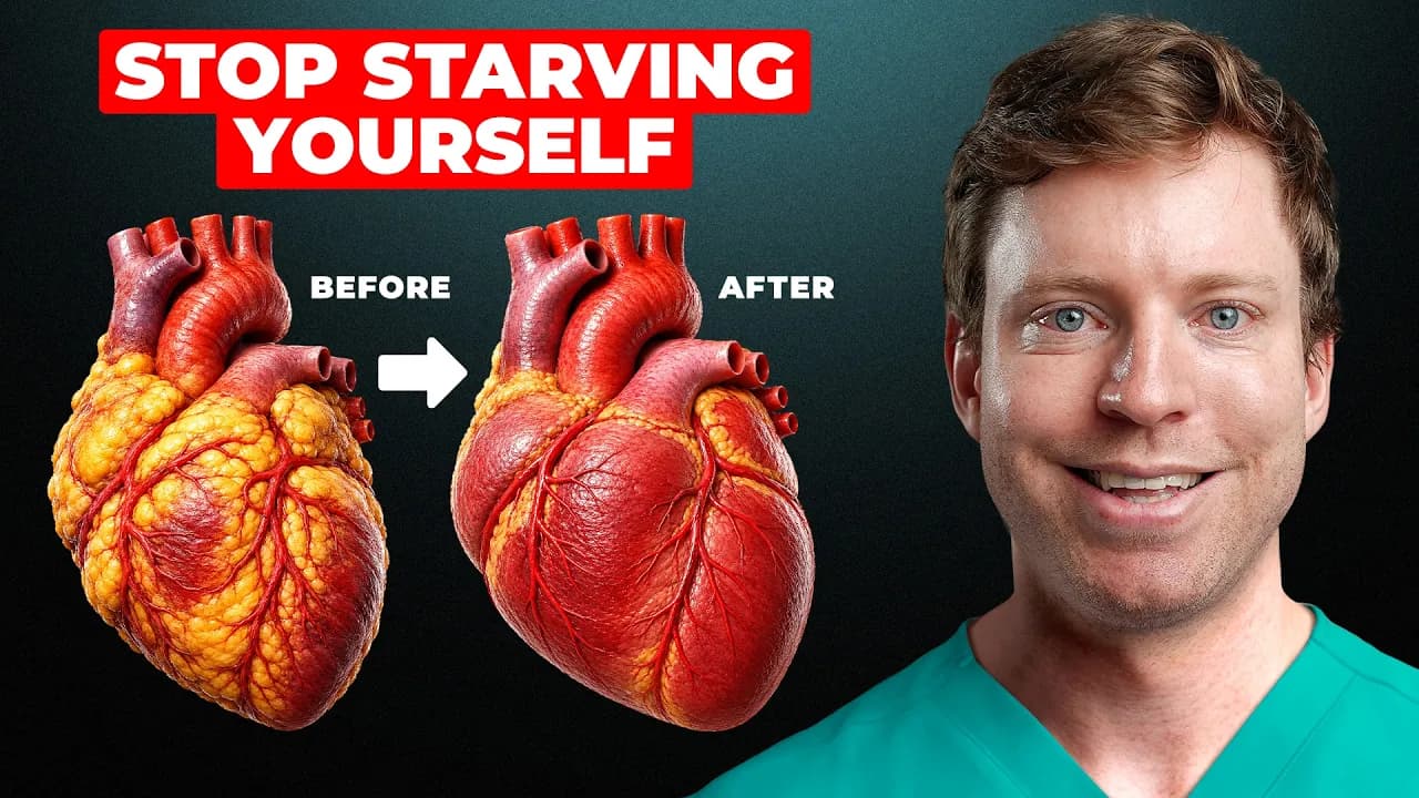 From Shrink the Fat Around Your Heart · video
See the scientific wording
Epicardial fat releases inflammatory molecules such as interleukin-6 and tumor necrosis factor alpha directly onto the heart muscle and coronary arteries, promoting cardiovascular disease.
Correlational — new studies may shift this
Observational4 moderate-quality studies link this claim to the outcome, but causation is not established.
What the research says
4 studies reviewedSupporting (4)
Correlation of Epicardial Fat Thickness With the Severity of Coronary Artery Disease
Cross-Sectional StudyHuman2025
People with more fat around their heart had much worse heart artery blockages, suggesting that this fat might be harming the heart — even though the study didn't measure the exact inflammatory chemicals.
Cross-Sectional StudyHuman2022
The study found that the fat surrounding the heart makes more inflammatory chemicals like IL-6 than other fat in the body, especially in people with heart disease. This supports the idea that this heart fat directly harms the heart and arteries by causing inflammation.
Cross-Sectional StudyHuman2024
This study found that the fat around the heart’s main arteries is more inflamed in people with a common heart rhythm problem, even when their arteries aren’t more blocked. This suggests that this fat is actively producing harmful inflammation that affects the heart.
Contradicting (0)
No contradicting studies found yet
That doesn't mean it's settled — it just means no study has tested the opposite.
Quality-weighted scoring: we follow the GRADE framework — each study is rated High, Moderate, Low, or Very Low based on study design, methodology rigor, and risk of bias. A single high-quality RCT can outweigh several weaker observational studies.
Scores reflect study quality, not just count.
Fat around the heart becomes stressed and inflamed, releasing high levels of interleukin-6 and tumor necrosis factor alpha directly onto the heart muscle and coronary arteries. These chemicals damage the inner lining of the arteries, cause smooth muscle cells to multiply abnormally, and attract immune cells that form plaque. At the same time, the fat stops producing protective molecules, making the damage worse. This process happens right where the fat touches the arteries and heart, leading to blockages and heart disease.
Score breakdown, mechanism chain, raw evidence, ideal studies needed & 4 supporting studies
How Fit Body Science checks a claim
- 1
We isolate the claim
Health advice from videos, articles and studies is broken down into single, testable claims.
- 2
We find the research
Each claim is matched against peer-reviewed studies, with every source cited by DOI.
- 3
We grade the evidence
Studies are scored on methodology, statistical rigor, transparency and publication quality.
The fitness and health internet is full of confident claims. We check them against real research.
Every claim on this site is traced back to peer-reviewed studies, scored on methodology and reporting quality, and given a verdict you can audit yourself — sources, DOIs and all.
- Full evidence breakdown and mechanism chains
- Ask our AI anything about a claim or its studies
- Get notified when new research changes a verdict
Epicardial fat produces inflammatory molecules including interleukin-6 and tumor necrosis factor alpha that act directly on the heart muscle and coronary arteries, contributing to the development of cardiovascular disease.
Mechanism
4 studiesFat around the heart gets stressed and starts pumping out harmful chemicals like interleukin-6 and tumor necrosis factor alpha. These chemicals attack the heart's blood vessels, making them narrow and stiff while stopping the production of protective signals. This directly causes plaque buildup and heart disease.
Fat around the heart becomes stressed and inflamed, releasing high levels of interleukin-6 and tumor necrosis factor alpha directly onto the heart muscle and coronary arteries. These chemicals damage the inner lining of the arteries, cause smooth muscle cells to multiply abnormally, and attract immune cells that form plaque. At the same time, the fat stops producing protective molecules, making the damage worse. This process happens right where the fat touches the arteries and heart, leading to blockages and heart disease.
Epicardial adipose tissue expands due to metabolic stress or reduced blood flow, leading to adipocyte hypoxia and dysfunction.
Dysfunctional adipocytes in epicardial fat downregulate adiponectin secretion and upregulate interleukin-6 and tumor necrosis factor alpha production.
Interleukin-6 and tumor necrosis factor alpha diffuse locally to adjacent coronary endothelial cells and vascular smooth muscle cells, inducing endothelial dysfunction and smooth muscle proliferation.
Chronic exposure to these cytokines promotes macrophage infiltration, oxidative stress, and plaque formation in coronary arteries.
Reduced adiponectin removes a key anti-inflammatory and vasoprotective signal, accelerating atherosclerosis and arterial stenosis.
Inflammatory mediators from pericoronary fat alter tissue composition, increasing water content and reducing lipid content, which amplifies local cytokine release and propagates damage to adjacent myocardial tissue.
Evidence from Studies
Last searched 2mo ago
Supporting (4)
Community contributions welcome
Correlation of Epicardial Fat Thickness With the Severity of Coronary Artery Disease
People with more fat around their heart had much worse heart artery blockages, suggesting that this fat might be harming the heart — even though the study didn't measure the exact inflammatory chemicals.
Relationship between Epicardial and Coronary Adipose Tissue and the Expression of Adiponectin, Leptin, and Interleukin 6 in Patients with Coronary Artery Disease
The study found that the fat surrounding the heart makes more inflammatory chemicals like IL-6 than other fat in the body, especially in people with heart disease. This supports the idea that this heart fat directly harms the heart and arteries by causing inflammation.
CT-Assessment of Epicardial Fat Identifies Increased Inflammation at the Level of the Left Coronary Circulation in Patients with Atrial Fibrillation
This study found that the fat around the heart’s main arteries is more inflamed in people with a common heart rhythm problem, even when their arteries aren’t more blocked. This suggests that this fat is actively producing harmful inflammation that affects the heart.
The study found that areas of the heart with more calcium buildup (a sign of artery disease) also had more inflammation in the surrounding fat, suggesting that this fat is actively contributing to heart disease.
Contradicting (0)
Community contributions welcome
Score Breakdown
No multi-axis breakdown available yet. The overall Pro / Against score above is the best signal.
- No clinical evidence is available; the score reflects mechanistic plausibility only.
What Would Prove This
Per GRADE and EBM methodology, here is what ideal scientific evidence would look like to definitively prove or disprove this claim, ordered from strongest to weakest.
Systematic Review of Epicardial Fat Volume and Inflammatory Marker Levels in Patients with Cardiovascular Disease
Population: Adults with and without cardiovascular disease; Intervention: Measurement of epicardial fat volume and serum levels of interleukin-6 and tumor necrosis factor alpha; Comparator: Low vs high epicardial fat groups; Outcome: Incidence and severity of cardiovascular disease; Duration: Longitudinal follow-up across included studies.
Randomized Trial of Epicardial Fat Reduction via Pharmacologic Intervention on Inflammatory Markers and Coronary Endothelial Function
Population: Adults with elevated epicardial fat and subclinical cardiovascular disease; Intervention: Pharmacologic agent targeting epicardial fat inflammation; Comparator: Placebo; Outcome: Changes in interleukin-6, tumor necrosis factor alpha, and coronary endothelial function; Duration: 12 months.
Prospective Cohort Study of Epicardial Fat Volume, Inflammatory Biomarkers, and Incident Cardiovascular Events
Population: Healthy adults aged 40–70; Intervention: None (observational); Comparator: Stratified by epicardial fat volume and inflammatory marker levels; Outcome: Incidence of myocardial infarction, stroke, or coronary revascularization; Duration: 10 years.
In Vitro Model of Epicardial Fat Adipocyte Secretion on Coronary Endothelial and Cardiomyocyte Inflammatory Response
Population: Human epicardial adipose tissue explants and primary coronary endothelial cells and cardiomyocytes; Intervention: Exposure to conditioned media from epicardial fat; Comparator: Control media; Outcome: Expression of interleukin-6 and tumor necrosis factor alpha in target cells; Duration: 24–72 hours.
Mouse Model of Epicardial Fat Expansion and Direct Assessment of Myocardial Inflammation and Coronary Dysfunction
Population: Genetically modified mice with inducible epicardial fat expansion; Intervention: Induction of epicardial fat hypertrophy; Comparator: Control mice without fat expansion; Outcome: Myocardial cytokine levels, coronary artery stiffness, and cardiac function; Duration: 8–16 weeks.