The thymus shrinks with age because supportive signals like IGF-1 and FGF-21 drop, the tissue turns to fat, and inflammation damages it.
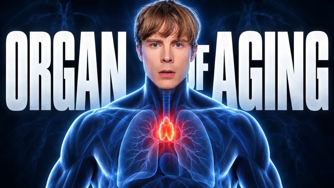 From The Organ That Controls How Fast You Age · video
See the scientific wording
Age-related thymic involution is caused by a decline in thymic support cells such as IGF-1 and FGF-21, disorganization of thymic architecture with replacement by fat, and damage to thymic tissue by increased pro-inflammatory cytokines; no effect sizes, absolute/relative risks, dosages, or durations are reported.
Correlational — new studies may shift this
Observational3 low-scoring studies link this claim to the outcome, but causation is not established.
What the research says
3 studies reviewedSupporting (3)
Cross-Sectional StudyHuman2025
This study shows that leaky guts and inflammation can make the thymus shrink with age, which fits part of the claim. But it doesn't mention IGF-1 or FGF-21, so those specific support-cell causes aren't tested here.
Cohort StudyAnimal2016
This mouse study found that a hormone called FGF21 drops with age and that boosting it keeps the thymus healthier and less fatty, while removing it speeds up aging. It supports part of the claim about FGF21 and fat, but didn't test IGF-1 or inflammation.
Enhanced paracrine action of FGF21 in stromal cells delays thymic aging
Cohort StudyAnimal2025
The study shows that boosting FGF21 in the thymus helps keep it young, and blocking FGF21's receptor makes aging worse, so low FGF21 likely contributes to thymus shrinkage. But the study didn't test IGF-1 or inflammation, so those parts of the claim are not proven here.
Contradicting (0)
No contradicting studies found yet
That doesn't mean it's settled — it just means no study has tested the opposite.
Quality-weighted scoring: we follow the GRADE framework — each study is rated High, Moderate, Low, or Very Low based on study design, methodology rigor, and risk of bias. A single high-quality RCT can outweigh several weaker observational studies.
Scores reflect study quality, not just count.
As the body ages, the thymus stops making enough of a hormone called FGF21. FGF21 normally tells the thymus support cells to keep working and to use fat for energy. Without enough FGF21, support cells lose their ability to help new T cells grow, fat builds up inside the thymus, and inflammatory signals from a leaky gut damage the thymus. The thymus shrinks, fills with fat, and makes fewer new T cells.
Score breakdown, mechanism chain, raw evidence, ideal studies needed & 3 supporting studies
Related videos
How Fit Body Science checks a claim
- 1
We isolate the claim
Health advice from videos, articles and studies is broken down into single, testable claims.
- 2
We find the research
Each claim is matched against peer-reviewed studies, with every source cited by DOI.
- 3
We grade the evidence
Studies are scored on methodology, statistical rigor, transparency and publication quality.
The fitness and health internet is full of confident claims. We check them against real research.
Every claim on this site is traced back to peer-reviewed studies, scored on methodology and reporting quality, and given a verdict you can audit yourself — sources, DOIs and all.
- Full evidence breakdown and mechanism chains
- Ask our AI anything about a claim or its studies
- Get notified when new research changes a verdict
The thymus shrinks with age because supportive signals like IGF-1 and FGF-21 drop, the tissue turns to fat, and inflammation damages it.
Mechanism
3 studiesThe thymus shrinks with age because it loses a hormone called FGF21 and gets hit by inflammation from a leaky gut. FGF21 normally keeps thymus support cells working and stops fat from building up. When FGF21 drops, support cells fail, fat fills the thymus, and inflammatory signals damage the tissue, so fewer new T cells are made.
As the body ages, the thymus stops making enough of a hormone called FGF21. FGF21 normally tells the thymus support cells to keep working and to use fat for energy. Without enough FGF21, support cells lose their ability to help new T cells grow, fat builds up inside the thymus, and inflammatory signals from a leaky gut damage the thymus. The thymus shrinks, fills with fat, and makes fewer new T cells.
Thymic stromal cells produce FGF21, and thymic FGF21 expression declines with age.
FGF21 binds βKlotho–FGFR1c complexes on cortical thymic epithelial cells and other thymic stromal cells, activating ERK phosphorylation.
ERK signaling maintains cortical thymic epithelial cell function and expression of thymopoietic factors Eva, Il7, and Fgf7, preserving earliest thymocyte progenitors and thymopoiesis.
Age-related loss of FGF21 signaling reduces lipid utilization and brown adipose tissue, causing ectopic intrathymic lipid accumulation and adipocyte emergence.
Ectopic lipid generates ceramides and other damage-associated molecular patterns, activating NLRP3 inflammasomes in thymic macrophages and increasing local inflammation.
Age-related loss of intestinal barrier integrity increases microbial translocation, raising systemic pro-inflammatory cytokines such as TNF-α and IL-6.
Pro-inflammatory cytokines damage thymic epithelial cells and stromal architecture, impairing thymic support function.
Combined stromal dysfunction, lipid accumulation, and inflammatory damage disorganize thymic architecture, replace thymic tissue with fat, reduce thymic T-cell export, and lower naive T-cell frequencies.
Evidence from Studies
Last searched 13d ago
Supporting (3)
Community contributions welcome
Age‐related loss of intestinal barrier integrity plays an integral role in thymic involution and T cell ageing
This study shows that leaky guts and inflammation can make the thymus shrink with age, which fits part of the claim. But it doesn't mention IGF-1 or FGF-21, so those specific support-cell causes aren't tested here.
Prolongevity hormone FGF21 protects against immune senescence by delaying age-related thymic involution
This mouse study found that a hormone called FGF21 drops with age and that boosting it keeps the thymus healthier and less fatty, while removing it speeds up aging. It supports part of the claim about FGF21 and fat, but didn't test IGF-1 or inflammation.
Enhanced paracrine action of FGF21 in stromal cells delays thymic aging
The study shows that boosting FGF21 in the thymus helps keep it young, and blocking FGF21's receptor makes aging worse, so low FGF21 likely contributes to thymus shrinkage. But the study didn't test IGF-1 or inflammation, so those parts of the claim are not proven here.
Contradicting (0)
Community contributions welcome
Score Breakdown
No multi-axis breakdown available yet. The overall Pro / Against score above is the best signal.
- No clinical evidence is available; the score reflects mechanistic plausibility only.
What Would Prove This
Per GRADE and EBM methodology, here is what ideal scientific evidence would look like to definitively prove or disprove this claim, ordered from strongest to weakest.
Systematic Review of Longitudinal and Mechanistic Studies on Thymic Involution Drivers
Systematic search and meta-analysis of prospective cohort studies, animal models, and human histology studies that repeatedly measure thymic volume/function, circulating IGF-1 and FGF-21, thymic architecture/fat content, and inflammatory cytokines (e.g., IL-6, TNF-α) over aging; assess temporality and consistency.
Randomized Trial of IGF-1/FGF-21 or Anti-Inflammatory Intervention on Thymic Involution in Older Adults
Double-blind RCT in adults aged 65+ with age-related thymic involution; randomize to IGF-1 analog, FGF-21 analog, anti-cytokine therapy, or placebo; measure thymic volume by CT/MRI, naive T-cell output, thymic fat content, and inflammatory markers at baseline and 6–12 months.
Prospective Cohort of Thymic Volume, IGF-1/FGF-21, and Cytokines in Aging Adults
Longitudinal cohort of healthy adults aged 20–80; measure thymic volume/function by imaging and flow cytometry, serum IGF-1/FGF-21, inflammatory cytokines, and thymic fat fraction every 1–2 years for 5–10 years; correlate changes over time.
Cross-Sectional Comparison of Thymic Architecture and Biomarkers Across Age Groups
Cross-sectional study comparing thymic tissue (from surgery/autopsy) or imaging and blood biomarkers in age-stratified groups (e.g., 20–30, 40–50, 60–70, 80+); quantify IGF-1/FGF-21 expression, thymic fat, and cytokine levels.
Animal Model of Thymic Involution with IGF-1/FGF-21 Knockout and Cytokine Blockade
Aged mice or non-human primates with conditional knockout of IGF-1/FGF-21 signaling, induced thymic fat accumulation, or cytokine blockade (e.g., anti-IL-6/TNF-α); measure thymic size, architecture, fat content, and T-cell output compared with wild-type controls over lifespan.