The thymus shrinks after puberty: it is about 95% active tissue in newborns, 60% by age 25, and 10% or less by age 70, replaced by fat.
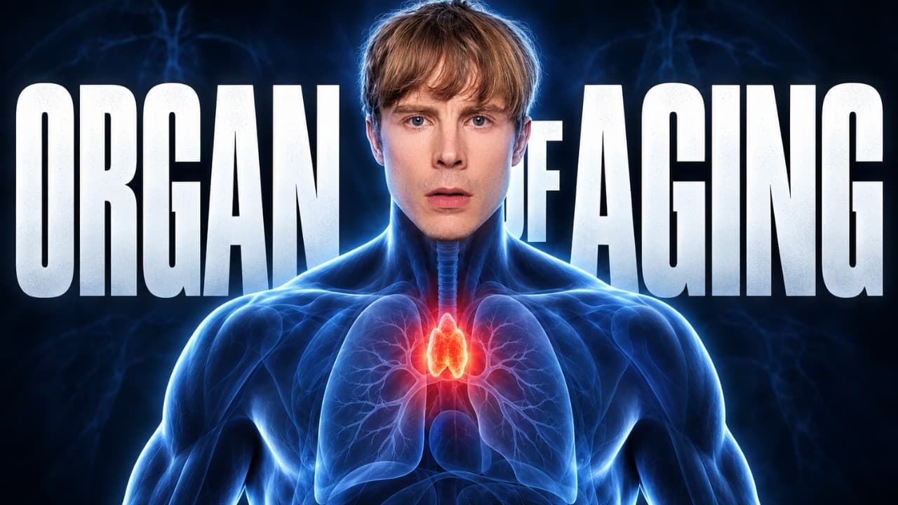 From The Organ That Controls How Fast You Age · video
See the scientific wording
Thymic involution begins after puberty and progresses with age, such that active thymic tissue is approximately 95% (ABSOLUTE proportion of thymic tissue) in newborns, about 60% (ABSOLUTE proportion) by age 25, and 10% or less (ABSOLUTE proportion) by around age 70; lost thymic tissue is replaced by fat and perivascular space. These are absolute age-specific proportions, not relative risks; no relative risk or likelihood is reported.
Correlational — new studies may shift this
Observational3 low-scoring studies link this claim to the outcome, but causation is not established.
What the research says
3 studies reviewedSupporting (3)
Cross-Sectional StudyHuman2025
The study shows that as the thymus ages, more fat cells build up, which matches the idea that the thymus shrinks and turns into fat. But it does not check the exact numbers in the claim, like 95% at birth or 10% by age 70.
Cross-Sectional StudyHuman2024
The study agrees that the thymus shrinks with age and turns into fat, but it does not give exact percentages for how much active tissue remains at different ages, so the specific numbers in the claim are not proven.
Multiscale physiologically-based model of age-dependent CD4+ T-lymphocyte homeostasis
Computational/Algorithm Study2026
The study agrees that the thymus gets weaker with age, but it doesn't give the specific percentages about how much tissue is left, so it can't confirm those exact numbers.
Contradicting (0)
No contradicting studies found yet
That doesn't mean it's settled — it just means no study has tested the opposite.
Quality-weighted scoring: we follow the GRADE framework — each study is rated High, Moderate, Low, or Very Low based on study design, methodology rigor, and risk of bias. A single high-quality RCT can outweigh several weaker observational studies.
Scores reflect study quality, not just count.
As people get older, the thymus makes more of a signal called thymosin-alpha1. This signal makes support cells in the thymus turn into fat cells. The fat builds up and takes the place of the working parts of the thymus. Because of this, the thymus makes fewer new immune cells over time. This is why the thymus shrinks and becomes mostly fat as people age.
Score breakdown, mechanism chain, raw evidence, ideal studies needed & 3 supporting studies
Related videos
How Fit Body Science checks a claim
- 1
We isolate the claim
Health advice from videos, articles and studies is broken down into single, testable claims.
- 2
We find the research
Each claim is matched against peer-reviewed studies, with every source cited by DOI.
- 3
We grade the evidence
Studies are scored on methodology, statistical rigor, transparency and publication quality.
The fitness and health internet is full of confident claims. We check them against real research.
Every claim on this site is traced back to peer-reviewed studies, scored on methodology and reporting quality, and given a verdict you can audit yourself — sources, DOIs and all.
- Full evidence breakdown and mechanism chains
- Ask our AI anything about a claim or its studies
- Get notified when new research changes a verdict
The thymus shrinks after puberty: it is about 95% active tissue in newborns, 60% by age 25, and 10% or less by age 70, replaced by fat.
Mechanism
2 studiesAs we age, the thymus makes more of a molecule called thymosin-alpha1. This molecule tells support cells in the thymus to become fat cells. The fat builds up and replaces the working parts of the thymus, so the thymus makes fewer new immune cells. This is why the thymus gets smaller and turns mostly to fat over time.
As people get older, the thymus makes more of a signal called thymosin-alpha1. This signal makes support cells in the thymus turn into fat cells. The fat builds up and takes the place of the working parts of the thymus. Because of this, the thymus makes fewer new immune cells over time. This is why the thymus shrinks and becomes mostly fat as people age.
Aging leads to increased production of thymosin-alpha1 within the thymus.
Thymosin-alpha1 stimulates thymic mesenchymal stromal cells, activating AKT phosphorylation and increasing FoxO1 expression and activity.
FoxO1 induces expression of MRAP in thymic mesenchymal stromal cells.
MRAP promotes adipogenic differentiation of thymic mesenchymal stromal cells, upregulating PPARγ, C/EBPα, and FABP4 and causing lipid accumulation.
Adipocytes accumulate in the thymus, replacing active thymic epithelial tissue and disrupting thymic architecture.
Reduced thymic epithelial space and altered stromal environment decrease thymocyte proliferation and differentiation, lowering production of mature CD4+ thymocytes.
Decreased egress of recent thymic emigrants into circulation reduces the supply of naive T cells.
Evidence from Studies
Last searched 13d ago
Supporting (3)
Community contributions welcome
MRAP mediated adipocyte differentiation by thymic mesenchymal stromal cells contributes to thymic involution
The study shows that as the thymus ages, more fat cells build up, which matches the idea that the thymus shrinks and turns into fat. But it does not check the exact numbers in the claim, like 95% at birth or 10% by age 70.
Transcriptional profile of human thymus reveals IGFBP5 is correlated with age-related thymic involution
The study agrees that the thymus shrinks with age and turns into fat, but it does not give exact percentages for how much active tissue remains at different ages, so the specific numbers in the claim are not proven.
Multiscale physiologically-based model of age-dependent CD4+ T-lymphocyte homeostasis
The study agrees that the thymus gets weaker with age, but it doesn't give the specific percentages about how much tissue is left, so it can't confirm those exact numbers.
Contradicting (0)
Community contributions welcome
Score Breakdown
No multi-axis breakdown available yet. The overall Pro / Against score above is the best signal.
Clinical support requires direct evidence. Mechanistic proxy and tangential studies contribute only to the mechanistic score.
- All linked studies are tangential or mechanistic proxies — no direct test of the claim has been found.
- No clinical evidence is available; the score reflects mechanistic plausibility only.
What Would Prove This
Per GRADE and EBM methodology, here is what ideal scientific evidence would look like to definitively prove or disprove this claim, ordered from strongest to weakest.
Systematic Review and Meta-Analysis of Age-Related Human Thymic Tissue Percentage
Systematic search of PubMed/Embase for human studies measuring active thymic tissue by CT, MRI, or histology from birth to >70 years; meta-analyze age-stratified proportions (newborns, ~25 years, ~70 years) and composition, with subgroup analyses by measurement modality, sex, and health status.
Longitudinal Cohort Study of Thymic Involution from Childhood to Older Age
Longitudinal cohort of healthy humans followed from pre-puberty through older adulthood with serial non-invasive thymic imaging (CT/MRI) every 5–10 years, measuring active thymic tissue volume/percentage and fat/perivascular replacement; outcomes compared across age, sex, and immune status.
Age-Stratified Cross-Sectional Study of Thymic Tissue Composition
Large cross-sectional study of individuals across ages (newborns, 25, 70) using CT/MRI or autopsy/histology to quantify active thymic tissue percentage and fat/perivascular space, with standardized age bins and measurement protocols.
Animal Model Study of Thymic Involution and Fat Replacement Across Lifespan
Longitudinal animal study (e.g., non-human primates or rodents) with serial thymic imaging and histology from birth to old age, measuring active thymic tissue percentage, fat infiltration, and perivascular space expansion.